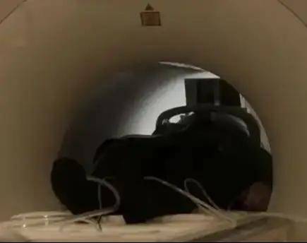
“I feel so uncomfortable, I am very scared, and I am a bit short of breath.”
Why did this subject experience such a situation? After the physician’s inquiries, it turned out that she had claustrophobia, and prior to the examination, she did not truthfully disclose her condition, leading to this issue.
So, what solutions are there for patients with claustrophobia undergoing PET/CT examinations?
Claustrophobia is an anxiety disorder related to enclosed spaces, a manifestation of phobia. It falls under the category of situational phobia, with patients fearing enclosed or crowded places such as elevators, train cars, or airplane cabins, worrying about unknown fears that may arise in these environments. Severe cases may even result in anxiety and obsessive symptoms, but once away from such environments, the patients’ physiological and behavioral responses return to normal.
With advancements and updates in medical equipment, the clinical application of PET/CT has become increasingly widespread, and its diagnostic value has been recognized clinically. However, patients with claustrophobia face various obstacles during the examination. Throughout the examination, patients must lie flat, with their entire bodies entering the cylindrical device, surrounded by walls, in a confined and dim space. Patients may feel oppressed and suffocated, making it challenging to remain still and cooperate to complete the examination. Additionally, the noise of the PET/CT machine may heighten the patients’ fears.
How can this problem be resolved? How can patients with claustrophobia undergo examinations normally?
Nurses in the nuclear medicine department should communicate gently with patients before the examination, listening carefully and patiently explaining the relevant knowledge about the examination, alleviating the patients’ anxiety and fear, and helping them relax before the examination.
Patients can be allowed to familiarize themselves with the resting area and the examination room in advance, creating a more humane examination atmosphere. The steps of the PET/CT whole-body tumor imaging process can be explained to the patients, and they can simulate the examination process before the actual procedure to minimize their fear.
When administering 18F-FDG to patients, clear explanations about the purpose of the injection should be provided to alleviate their concerns. If necessary, family members can accompany the patients into the examination room and comfort them physically to provide a sense of security.
Patients should be informed that they can communicate with the technician at any time during the examination, and the technician should maintain communication with the patients throughout the process, allowing them to feel the warmth of the medical staff. Affirmation and encouragement of the patients’ cooperation can be provided, along with sharing successful experiences of similar cases with them.
Patients should be taught how to use a call button, making them aware that they can always call for medical personnel if they feel unwell, enhancing their sense of safety; the lighting in the examination room should be adjusted to be soft, alleviating their fear, and the lights of the scanner should be turned on to create a sense of space.
Case Example
A 47-year-old male patient underwent PET/CT examination to search for tumor lesions due to elevated tumor markers. On the day of the examination, the admitting physician, while asking about the general medical history, learned that the patient had a fear of enclosed environments, having previously exited the MRI examination table on his own. The admitting physician then asked the patient to lie on the PET/CT examination table. As soon as the patient entered the circular machine, he attempted to climb out of the machine, expressing fear, panic, and shortness of breath, saying he could not endure it.
In response to this situation, the physician proposed two plans to ensure the examination proceeded smoothly:
Utilizing psychological methods to help the patient gradually adapt by administering anti-anxiety medication or sedatives.
After communicating with the patient and his family, the first plan was adopted, with the following process:
1) Instruct the patient to stretch his head into the circular machine frame, exiting if feeling uncomfortable, lasting about 5 minutes;
2) Ask the patient to lie on the examination table, while the physician controls the table, slowly bringing him in and out of the circular scanner several times. The patient transitioned from resisting strongly to gradually adapting, over a period of approximately 10 minutes;
3) The patient reported that once he entered the frame…


